CASE REPORT
Malignant Phyllodes Tumor of the Breast: Case Report
Tumor Filoide Maligno de Mama: Relato de Caso
Tumor Phyllodes Maligno de Mama: Reporte de Caso
doi: https://doi.org/10.32635/2176-9745.RBC.2022v68n3.2568
Rafael Everton Assunção Ribeiro da Costa1; Luis Felipe Rodrigues Brandão de Barros2; Raimundo Gerônimo da Silva Júnior3; Marcos Antonio Veras de Negreiros4; Eid Gonçalves Coelho5; Antonio Luiz Moreira Junior6; Carlos Eduardo Coelho de Sá7
1-3Universidade Estadual do Piauí, Centro de Ciências da Saúde. Teresina (PI), Brazil. E-mails: rafaelearcosta@gmail.com; luisbarros@aluno.uespi.br; gerjrpi@gmail.com. Orcid iD: https://orcid.org/0000-0002-0798-890X; Orcid iD: https://orcid.org/0000-0002-1690-6638; Orcid iD: https://orcid.org/0000-0003-4032-6710
4-7Unidade de Assistência de Alta Complexidade em Oncologia, Hospital Macrorregional de Caxias Dr. Everardo Aragão. Caxias (MA), Brazil. E-mails: marcosnegreiros@hotmail.com; eidgcoelho@hotmail.com; antoniolmoreirajr@gmail.com; eduardo_sa1601@hotmail.com. Orcid iD: https://orcid.org/0000-0002-6675-5559; Orcid iD: https://orcid.org/0000-0001-9955-3737; Orcid iD: https://orcid.org/0000-0002-8985-1586; Orcid iD: https://orcid.org/0000-0002-9610-8010
Corresponding author: Rafael Everton Assunção Ribeiro da Costa. Rua Olavo Bilac, 2335 – Centro. Teresina (PI), Brazil. E-mail: rafaelearcosta@gmail.com
ABSTRACT
Introduction: Phyllodes tumors (PT) are rare and account for 0.3% to 0.5% of all breast tumors. PT may be classified as benign, borderline or malignant. The aim of this study was to report a case of malignant PT of the breast. Case report: A 27-year-old woman presented with a mass in the left breast with histopathological features of malignancy (results of US of the breast: an oval, lobulated hypoechogenic lesion, measuring 7.7 cm – BI-RADS® 4C). A segmental resection (SR) of the breast was performed and histopathology study of the surgical specimen confirmed a malignant PT. Adjuvant radiotherapy was used for supplemental treatment. One year later, the patient had a local recurrence of the primary tumor and underwent a new SR of the left breast. There was no indication of breast reirradiation. At about 31 months after diagnosis (September 2019 – April 2022), the patient is well and adherent to periodical clinical follow-up. Conclusion: This study presents a case of malignant PT that occurred in a young patient and had a more aggressive course.
Key words: breast neoplasms; phyllodes tumor; mastectomy, segmental; radiotherapy, adjuvant; case reports.
RESUMO
Introdução: Os tumores filoides (TF) são raros e representam entre 0,3% e 0,5% dos tumores de mama, podendo ser classificados como benignos, borderline ou malignos. O objetivo deste estudo foi relatar um caso de TF maligno de mama. Relato do caso: Mulher, 27 anos de idade, apresentando nódulo em mama esquerda com características histopatológicas de malignidade (resultados da ultrassonografia de mamas: lesão hipoecogênica, oval e lobulada, com 7,7 cm – BI-RADS® 4C). Foi realizada ressecção segmentar (RS) da mama e o histopatológico da peça cirúrgica mostrou tratar-se de um TF maligno. Como tratamento complementar, foi realizado radioterapia adjuvante. A paciente apresentou recidiva local do tumor primário em cerca de apenas um ano, sendo realizada nova RS da mama esquerda. Não houve indicação de reirradiação da mama. Em 31 meses após o diagnóstico (setembro de 2019 – abril de 2022), a paciente encontra-se em bom estado geral e realizando seguimento clínico periódico. Conclusão: Este estudo apresenta um caso de TF maligno que ocorreu em uma paciente jovem e teve um curso mais agressivo.
Palavras-chave: neoplasias da mama; tumor filoide; mastectomia segmentar; radioterapia adjuvante; relatos de casos.
RESUMEN
Introducción: Los tumores phyllodes (TP) son poco frecuentes y representan del 0,3% al 0,5% de todos los tumores de mama. Los TP pueden clasificarse como benigno, limítrofe o maligno. El objetivo de este estudio fue reportar un caso de TP maligno de mama. Reporte del caso: Una mujer de 27 años se presentó con una masa en la mama izquierda con características histopatológicas de malignidad (resultados de la ecografía de mama: lesión hipoecogénica ovalada y lobulada, de 7,7 cm – BI-RADS® 4C). Se realizó una resección segmentaria (RS) de la mama y el estudio histopatológico de la pieza quirúrgica confirmó un TP maligno. Se utilizó radioterapia adyuvante como tratamiento complementario. Un año después, la paciente presentó una recidiva local del tumor primario y fue sometida a una nueva RS de mama izquierda. No hubo indicación de reirradiación mamaria. Aproximadamente 31 meses después del diagnóstico (septiembre de 2019 – abril de 2022), la paciente se encuentra bien y se adhiere al seguimiento clínico periódico. Conclusión: Este estudio presenta un caso de TP maligno que ocurrió en una paciente joven y tuvo un curso más agresivo.
Palabras clave: neoplasias de la mama; tumor filoide; mastectomía segmentaria; radioterapia ayuvante; informes de casos.
INTRODUCTION
Phyllodes tumor (PT) is a rare breast neoplasm. The incidence of PT is 0.3-0.5% of all breast tumors, occurring most commonly in women aged 35 to 65. PT has a benign origin in around 85-90% of the cases1. They are classically divided into benign, borderline or malignant2. Patients with PT usually present a fast-growing palpable mass with features similar to fibroadenoma on ultrasonography (US)3. Local recurrence of malignant PT is approximately 19-40%, and the mean period of recurrence is 1-39 months (mean: 20.3 months). The risk of distant metastases is higher than 75% in the first 120 months, meaning a worse prognosis4.
The treatment of choice for these tumors is segmental resection (SR) of the breast with adequate margins1. Adjuvant radiotherapy is also indicated following SR in cases of borderline or malignant tumors larger than 5.0 cm and/or involved margins2. Adjuvant radiotherapy has been shown to increase disease-free survival in these cases. However, available data on increased overall survival in the literature remain inconclusive3. The aim of this study was to report a case of malignant PT of the breast.
The Institutional Review Board of “Universidade Estadual do Piauí”, Teresina (PI), Brazil – reference number 5.137.868 approved the study. The patient signed the informed consent form (ICF).
CASE REPORT
A 27-year-old woman sought medical attention in April 2019 presenting with a lump in the left upper outer quadrant (LUOQ) of the left breast – measuring 9 cm on physical examination. US of the breast revealed an oval, lobulated hypoechogenic lesion, measuring 7.7 cm (BI-RADS® 4C). Core needle biopsy and histopathological study demonstrated a lesion with spindle cell proliferation and discrete atypia. Further investigation of the surgical specimen was indicated and SR of the breast was performed in August 2019 (Figure 1).
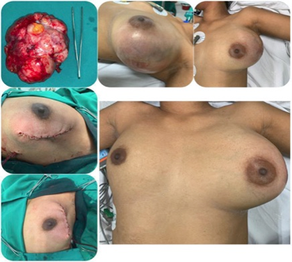
Figure 1. Segmental resection for treatment of the primary tumor
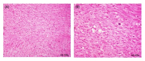
In September 2019, histopathology study of the surgical specimen (Figure 2) showed a fibroepithelial tumor with the following features: areas of stromal expansion, spindle cell proliferation, atypias and frequent mitotic figures – with clear surgical margins. 5 months after initial symptoms, the definitive diagnosis was malignant PT. From February to April 2020, the patient underwent 3D adjuvant radiotherapy. The dose delivered was 50 Gy in 25 fractions to the left breast + boost of 10 Gy in 5 fractions to the LUOQ.
|
|
|
Figure 2.
Histopathology of the surgical specimen (first segmental resection) –
hematoxylin-eosin (H&E). (A). At 100x magnification. |
In September 2020, there was a recurrence of the primary tumor of the left breast (Figure 3), with detection of a mass measuring 5.5 cm by a new US of the breast. The patient underwent a new surgical resection. Adjuvant reirradiation was not indicated. At about 31 months after diagnosis (September 2019 – April 2022), she is well and is adherent to periodical clinical follow-up at the medical unit.
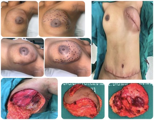
Figure 3. Segmental resection for treatment of local recurrence
DISCUSSION
PT is a rare form of breast tumor. It accounts for about 2.5% of fibroepithelial tumors of the breast2, with an incidence of 2.1 per million women3. PT of the breast usually manifests as a painless rapidly growing unifocal mass that is most commonly located in the LUQ, as occurred in the case report4. The patient developed a malignant PT before 30 years of age, which is a major rare event.
The pathogenesis of PT is currently unclear. However, it is known that it is related to diverse factors, including hormone disorders (primarily hyperestrogenism), breast injury, ethnicity and breastfeeding. It may also occur during pregnancy1,4. Histologically, PT is characterized by a double-layered leaf-like epithelial pattern with hypercellular stroma2.
The treatment of choice for these cases is SR with a 1.0 cm margin and without axillary node dissection1-5. According to a systematic review with meta-analysis conducted by Toussaint et al., the width of the safety margin is related to lower local recurrence, mainly in malignant PT, although a significant difference in the occurrence of metastases was not found6. In the present case, the primary tumor was treated with SR and left breast irradiation. However, about one year later, there was a local recurrence with repeated SR.
Radiotherapy has been shown to be effective at preventing local recurrence, in addition to increasing functioning of the area and patient satisfaction2. In general, 45-50 Gy is delivered to subclinical lesions1. For this patient, a dose 50 Gy in 25 fractions to the primary tumor of the left breast + boost of 10 Gy in 5 fractions to the LUOQ were used. Left breast reirradiation was not indicated.
Due to limited data on chemotherapy for PT of the breast, systemic chemotherapy is not indicated in these cases, except in non-resectable distant metastases (uncommon event)7,8. This modality of treatment was not indicated for this patient.
At about 31 months after diagnosis, the patient is in good clinical condition and adheres to periodical clinical follow-up in the medical unit.
CONCLUSION
This study presents a case of malignant PT that occurred in a young patient and had a more aggressive course.
Rafael Everton Assunção Ribeiro da Costa, and Luis Felipe Rodrigues Brandão de Barros: Study design, data acquisition, quality control of data, data analysis and interpretation, wording and review. Raimundo Gerônimo da Silva Júnior, Marcos Antonio Veras de Negreiros, Eid Gonçalves Coelho, Antonio Luiz Moreira Junior, and Carlos Eduardo Coelho de Sá: Study design, acquisition, quality control, analysis and interpretation of the data, wording, and critical review. All the authors approved the final version to be published.
DECLARATION OF CONFLICT OF INTERESTS
There is no conflict of interests to declare.
FUNDING SOURCES
None.
REFERENCES
1. Wu H, Li L, Yang J, et al. Radiotherapy with apatinib for recurrence of malignant phyllodes tumor of the breast: a case report. Medicine (Baltimore). 2020;99(3):e18808. doi: https://doi.org/10.1097/MD.0000000000018808
2. Liu HP, Chang WY, Hsu CW, et al. A giant malignant phyllodes tumor of breast post mastectomy with metastasis to stomach manifesting as anemia: a case report and review of literature. BMC Surg. 2020;20(1):187. doi: https://doi.org/10.1186/s12893-020-00846-0
3. Koukourakis IM, Zygogianni A, Kouloulias V, et al. Successful treatment of a locally recurrent and metastatic malignant phyllodes tumor with accelerated radiotherapy and Nab-paclitaxel, cisplatin, and liposomal doxorubicin chemotherapy. Chemotherapy. 2021;66(3):82-6. doi: https://doi.org/10.1159/000517246
4. Fang CL, Hsu CH, Tu CW. Malignant phyllodes tumor recurrence in the pleural cavity via the deep inferior epigastric perforator flap and internal mammary vessel bundle: a case report. Ann Plast Surg. 2019;82(6):618-21. doi: https://doi.org/10.1097/SAP.0000000000001795
5. Hasdemir S, Tolunay Ş, Özşen M, et al. Phyllodes tumor of the breast: a clinicopathological evaluation of 55 cases. Eur J Breast Health. 2020;16(1):32-8. doi: https://doi.org/10.5152/ejbh.2019.4709
6. Toussaint A, Piaget-Rossel R, Stormacq C, et al. Width of margins in phyllodes tumors of the breast: the controversy drags on?-a systematic review and meta-analysis. Breast Cancer Res Treat. 2021;185(1):21-37. doi: https://doi.org/10.1007/s10549-020-05924-8
7. Roberts N, Runk DM. Aggressive malignant phyllodes tumor. Int J Surg Case Rep. 2015;8:161-5. doi: https://doi.org/10.1016/j.ijscr.2014.12.041
8. Fernández-Ferreira R, Arroyave-Ramírez A, Motola-Kuba D, et al. Giant benign mammary phyllodes tumor: report of a case and review of the literature. Case Rep Oncol. 2021;14(1):123-33. doi: https://doi.org/10.1159/000510741
Recebido em 2/2/2022
Aprovado em 26/4/2022
Scientific-Editor: Anke Bergmann. Orcid iD: https://orcid.org/0000-0002-1972-8777
![]()
Este é um artigo publicado em acesso aberto (Open Access) sob a licença Creative Commons Attribution, que permite uso, distribuição e reprodução em qualquer meio, sem restrições, desde que o trabalho original seja corretamente citado.
©2019 Revista Brasileira de Cancerologia | Instituto Nacional de Câncer José Alencar Gomes da Silva | Ministério da Saúde