CASE REPORT
Intraoral Stent for Head and Neck Radiotherapy in Patients with Palate Tumor
Moldeira Intraoral para Radioterapia de Cabeça e Pescoço em Pacientes com Tumor de Palato
Stent Intraoral para Radioterapia de Cabeza y Cuello en Pacientes con Tumores Palatinos
https://doi.org/10.32635/2176-9745.RBC.2026v72n3.5476
Lísia Daltro Borges Alves1; Marco Túlio Cunha Santos2; Ana Carolina dos Santos Menezes3; Luciana Ferreira Stahel Lage4; Débora Lima Pereira5; Felipe Erlich6; Fernando Luiz Dias7; Héliton Spíndola Antunes8
1,3,8Instituto Nacional do Câncer (INCA), Divisão de Pesquisa Clínica e Desenvolvimento Tecnológico. Rio de Janeiro (RJ), Brasil. E-mails: lisia_94@hotmail.com; ana.smenezes@gmail.com; helitonspindola@gmail.com. Orcid iD: https://orcid.org/0000-0001-6779-8692; Orcid iD: https://orcid.org/0000-0001-6622-4400; Orcid iD: https://orcid.org/0000-0002-1076-8019
2INCA, Serviço de Radioterapia, Residência Multiprofissional em Oncologia. Rio de Janeiro (RJ), Brasil. E-mail: marcotuliocs@gmail.com. Orcid iD: https://orcid.org/0000-0002-2654-7392
4,5INCA, Serviço de Odontologia. Rio de Janeiro (RJ), Brasil. E-mails: lustahel@gmail.com; debora.lpereira@gmail.com. Orcid iD: https://orcid.org/0000-0002-2528-8579; Orcid iD: https://orcid.org/0009-0006-7965-6679
6INCA, Serviço de Radioterapia. Rio de Janeiro (RJ), Brasil. E-mail: ferlich@inca.gov.br. Orcid iD: https://orcid.org/0000-0003-4433-4208
7INCA, Serviço de Cirurgia de Cabeça e Pescoço. Rio de Janeiro (RJ), Brasil. E-mail: fdias@inca.gov.br. Orcid iD: https://orcid.org/0000-0003-1000-7436
Corresponding author: Lísia Daltro Borges Alves. INCA, Divisão de Pesquisa Clínica e Desenvolvimento Tecnológico. Rua André Cavalcanti, 37 – Centro. Rio de Janeiro (RJ), Brasil. CEP 20231-050. E-mail: lisia_94@hotmail.com
ABSTRACT
Introduction: Intraoral stents are devices applied to minimize the adverse effects of radiotherapy for head and neck tumors. This study aims to report the use of the intraoral stent in two cases of palate tumor treated with surgery and adjuvant radiotherapy. Case report: The first case refers to a 26-year-old man with mucoepidermoid carcinoma; the second case is a 55-year-old woman with cystic adenoid carcinoma. Before radiotherapy, an intraoral stent was made for both patients, composed of two acetate plates (upper/lower) and, in the lower, a plateau to down-grade and stabilize the tongue was made. Two columns of acrylic resin were used as constant height occlusal stops. The device was used during planning computer tomography and in all treatment sessions of radiotherapy. The plans were evaluated for medium doses, and low doses were observed in the mandible and parotid glands. Oral mucositis lesions were restricted to the region of the tumor, with maximum grades 2 and 3, respectively, only in the last 10 fractions of radiotherapy. After more than five years of follow-up, none of the patients were diagnosed with chronic toxicities associated with radiotherapy, such as xerostomia or osteoradionecrosis. Conclusion: These devices highlight the relevance of interdisciplinary planning and directly contribute to a better quality of life during and after radiotherapy for patients with tumors of the palate.
Key words: Head and Neck Neoplasms/drug therapy; Radiotherapy/adverse effects; Palate/surgery; Radiation Protection/instrumentation.
RESUMO
Introdução: Os stents intraorais são dispositivos aplicados para minimizar os efeitos adversos da radioterapia em tumores de cabeça e pescoço. Este estudo tem como objetivo relatar o uso do stent intraoral em dois casos de tumor de palato, tratados com cirurgia e radioterapia adjuvante. Relato dos casos: O primeiro caso refere-se a um homem de 26 anos com carcinoma mucoepidermoide; o segundo, a uma mulher de 55 anos com carcinoma adenoide cístico. Previamente à radioterapia, foi confeccionado um stent intraoral para ambos os pacientes, composto por duas placas de acetato (superior/inferior) e, na inferior, um platô para rebaixamento e estabilização da língua. Duas colunas de resina acrílica foram utilizadas como batentes oclusais de altura constante. O dispositivo foi utilizado durante a tomografia computadorizada de planejamento e em todas as sessões de tratamento radioterápico. Os planejamentos foram avaliados para doses médias, apresentando doses consideradas baixas na mandíbula e nas glândulas parótidas. As lesões de mucosite oral restringiram-se à região do tumor, com graus máximos 2 e 3, respectivamente, apenas nas últimas dez frações de radioterapia. Após mais de cinco anos de acompanhamento, nenhum dos pacientes foi diagnosticado com toxicidades crônicas associadas à radioterapia, como xerostomia ou osteorradionecrose. Conclusão: Esses dispositivos ressaltam a relevância do planejamento interdisciplinar e contribuem diretamente para uma melhor qualidade de vida durante e após a radioterapia em pacientes com tumores de palato.
Palavras-chave: Neoplasias de Cabeça e Pescoço/tratamento farmacológico; Radioterapia/efeitos adversos; Palato/cirurgia; Proteção Radiológica/instrumentação.
RESUMEN
Introducción: Los stents intraorales son dispositivos que se utilizan para minimizar los efectos adversos de la radioterapia en tumores de cabeza y cuello. Este estudio tiene como objetivo informar el uso del stent intraoral en dos casos de tumor de paladar, tratados con cirugía y radioterapia adyuvante. Informe de los casos: El primer caso se refiere a un hombre de 26 años con carcinoma mucoepidermoide; el segundo, es una mujer de 55 años con carcinoma adenoide quístico. Previo a la radioterapia, se fabricó un stent intraoral para ambos pacientes, compuesto por dos placas de acetato (superior/inferior) y, en la inferior, se realizó una plancha para rebajar y estabilizar la lengua. Se utilizaron dos columnas de resina acrílica como topes oclusales de altura constante. El dispositivo se utilizó durante la tomografía computarizada de planificación y en todas las sesiones de tratamiento de radioterapia. Las planificaciones se evaluaron para dosis medias, y se observaron dosis bajas en la mandíbula y las glándulas parótidas. Las lesiones de mucositis oral se limitaron a la región tumoral, con grados máximos 2 y 3, respectivamente, solo en las últimas diez fracciones de radioterapia. Después de más de cinco años de seguimiento, ninguno de los pacientes fue diagnosticado de toxicidades crónicas asociadas a la radioterapia, como xerostomía u osteorradionecrosis. Conclusión: Estos dispositivos resaltan la importancia de la planificación interdisciplinaria y contribuyen directamente a una mejor calidad de vida durante y después de la radioterapia en pacientes con tumores del paladar.
Palabras clave: Neoplasias de Cabeza y Cuello/tratamiento farmacológico; Radioterapia/efectos adversos; Hueso Paladar/cirugía; Protección Radiológica/ instrumentación.
INTRODUCTION
The incidence of oral cancer worldwide in 2018 was 246,420 cases for men and 108,444 for women1. Salivary gland tumors are considered a specific group and can affect the major (parotid, submandibular, and sublingual) or the minor salivary glands. In Brazil, these tumors' incidence ranges between 0.25% and 2.6%, and, especially when it comes to minor salivary glands, they are malignant in 28.9 to 57.7% of the cases2.
The most frequent malignant histological subtypes are mucoepidermoid carcinoma and adenoid cystic carcinoma2. The treatment is surgical resection, which may include adjuvant radiotherapy (RT)2. RT is considered an effective local therapeutic modality, but is associated with adverse effects like xerostomia, oral mucositis (OM), osteoradionecrosis (ORN), dysgeusia, trismus, and radiation-related caries3,4. These effects can impact the quality of life of patients, both during and after treatment. To minimize these adverse effects, an intraoral stent during RT sessions was proposed5-7.
The intraoral stent keeps the mouth open, and the tongue stabilized on the floor of the mouth8,9. Therefore, when using Intensity-Modulated Radiotherapy (IMRT) techniques, radiation can be directed to the tumor area without irradiating healthy structures10-13. Currently, few studies address this topic, especially tumors affecting the palate. Thus, this study aimed to report the use of the intraoral stent in two cases of palate tumor treated with surgery and adjuvant RT.
CASE REPORT
Two patients with a palate tumor were treated at the Instituto Nacional do Câncer, Brazil. Individual intraoral stents were made and used during the RT sessions. The patients were treated by IMRT, and the values of mean doses predicted in the mandible and bilateral parotids were analyzed, using the Eclipse-Varian treatment planning software.
The patients were followed daily by a specialized dental surgeon during the RT. OM was classified between grades 0 and 4, according to the World Health Organization (WHO) classification14. Due to the diagnosis of OM, they were enrolled in the low-power laser therapy protocol (100mW, 2J, 71.4J/cm², 0.028cm², emitting continuous light at 660nm), in daily applications, except for the tumor region.
These cases were approved by the Institutional Research Ethics Committee, report number 3,760,979 (CAAE (submission for ethical review): 25980019.2.0000.5274) and both patients signed the informed consent form, in compliance with Resolution 510/2016 of the National Health Council (CNS)15.
Case 1
A 26-year-old man with a palate mucoepidermoid carcinoma, T3N0M0, stage III, who underwent partial maxillectomy and adjuvant RT also involving the lymphatic chains IB and II (60Gy in 30 fractions). His intraoral stent was made with a 2.5cm mouth opening (Figures 1A and 1B). The mean doses to the mandible, right, and left parotid were respectively 36.78, 30.07, and 34.30Gy (Figure 1C). This case evolved with grade 2 OM in the right buccal mucosa and soft palate (20th to 25th sessions), treated with low-power laser therapy. There were no visible changes or complaints in the salivary condition during RT.
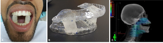
Figure 1. Case 1. A. Extraoral photograph with intraoral stent; B. Intraoral stent; C. Sagittal section of the radiotherapy planning system
Case 2
A 55-year-old woman with an adenoid cystic carcinoma in the hard and soft palate treated with partial maxillectomy associated with adjuvant RT excluding neck irradiation (60Gy in 30 fractions). Staging was not possible since the first surgical approach was performed at another hospital. Her intraoral stent was made with a 2.8cm mouth opening (Figures 2A and 2B). The mean doses to the mandible, right, and left parotid were respectively 10.06, 14.90, and 15.19Gy (Figure 2C). The patient presented OM grade 2 in the buccal mucosa, upper gingiva, hard palate, and upper vestibule, starting in the 22nd session, which evolved to grade 3 in the 24th, remaining until the 30th session, being treated with low-power laser therapy. There were also no complaints or visible changes in the salivary condition. The acetate sheets presented small cracks on two different occasions, requiring repair (gluing with acrylic resin at the crack sites) to ensure the treatment was not compromised.
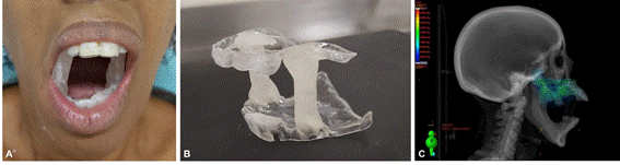
Figure 2. Case 2 A. Extraoral photograph with intraoral stent; B. Intraoral stent; C. Sagittal section of the radiotherapy planning system
Patients have been followed up for 5 years and 7 months and 5 years and 8 months respectively, with no evidence of disease, rehabilitated with obturator prostheses, without dietary restrictions or complaints related to phonation, xerostomia, radiation-related caries lesions or ORN.
DISCUSSION
Although there is no robust scientific evidence to indicate the use of the intraoral stents during head and neck cancer patients' RT, both cases presented benefits9. Currently, most studies have analyzed lower arch tumors12. In this context, few studies have investigated the use in maxillary tumors, and most of them are retrospective9,16-18.
Interestingly, both patients developed mucositis exclusively in the regions close to the tumor, leaving the opposite arch free from injuries. Furthermore, OM began on the twentieth session of RT, five days later than expected. These results are consistent with other studies, which reported a reduction in the OM incidence and severity5,6,18, and a delay in the OM onset when using the intraoral stent8. For palate tumors, the use of an intraoral stent can reduce acute and chronic xerostomia19, as noted in the reported cases.
The RT dose and fractionation will depend on the staging and compromise of the surgical margins and can reach up to 70Gy3. Both patients had a prescribed dose of 60Gy, and the mandibles received doses below 40Gy. The doses in case 2 are expressively lower when compared to case 1 due to lymph node irradiation. Doses above 50Gy in the jawbone are considered a risk factor for ORN, and intraoral stents can prevent ORN through dose reduction6,8.
Both patients used the intraoral stent during the RT with no significant complaints. Acrylic resin is usually the preferred material for handmade intraoral stents because it is non-toxic and easy to handle6,8. In the radiology service, acetate is used to make the upper and lower plates and acrylic resin for the pillars, with the mouth opening individually established for each patient. The intraoral stent can be made in a single consultation if the dentist is trained or if a prosthetic laboratory is available at the institution8,12,20.
The direct materials cost for making an intraoral stent ranges from 140 to 200 Brazilian reais20. When performed in a private service, the cost of the dental consultation and laboratory steps must be added, significantly increasing the associated cost. However, when used in public services (Brazilian National Health System – SUS), as in the cases presented, these devices become even more accessible and attractive, especially when considering toxicity reduction and treatment costs during and after RT11. Another option, especially in centers with greater investment in technology, is printing intraoral stents using 3D printers, but this requires a larger initial investment21.
The observed benefits highlight the importance of implementing the intraoral stent as a routine in large cancer centers. In addition, it reinforces the need for a multiprofessional team in oncology to minimize oral toxicities and consequently improve the quality of life of these patients. The clinical outcomes observed in these two cases are promising; however, multicenter randomized clinical trials are encouraged to standardize the use of this device.
CONCLUSION
The current cases presented OM only in tumoral regions and adjacent structures, with maximum grade 2 and 3, respectively. Small radiation doses in the mandible were also evident. After more than five years of follow-up, they didn’t present chronic RT toxicities.
ACKNOWLEDGEMENTS
To the multidisciplinary team, INCA patients, and Mônica Eliana Ribeiro Matos, prosthetics laboratory technician, for performing the laboratory work on intraoral stents.
CONTRIBUTIONS
Lísia Daltro Borges Alves, Marco Túlio Cunha Santos, Ana Carolina dos Santos Menezes, and Héliton Spíndola Antunes contributed substantially to the conception or planning of the study; Luciana Ferreira Stahel-Lage, Débora Lima Pereira, Felipe Erlich, and Fernando Luiz Dias contributed to the acquisition of the data; Lísia Daltro Borges Alves, Marco Túlio Cunha Santos, Ana Carolina dos Santos Menezes, and Héliton Spíndola Antunes participated in the analysis and/or interpretation of the data; all the authors participated in the writing and critical review and approved the final version for publication.
DECLARATION OF CONFLICT OF INTERESTS
There is no conflict of interests to declare.
DATA AVAILABILITY STATEMENT
All content underlying the text is contained in the manuscript.
FUNDING SOURCES
None.
REFERENCES
1. Bray F, Ferlay J, Soerjomataram I, et al. Global cancer statistics 2018: GLOBOCAN estimates of incidence and mortality worldwide for 36 cancers in 185 countries. CA Cancer J Clin. 2018;68:394-424. doi: https://doi.org/10.3322/caac.21492
2. Sterner F, Högmo A, Tano K. Carcinomas of the minor salivary glands of the oral cavity. A population-based study from the Swedish Head and neck cancer register for 2008-2018. Acta Otolaryngol. 2023;143(4):340-5. doi: https://doi.org/10.1080/00016489.2023.2191646
3. Jham BC, Reis PM, Miranda EL, et al. Oral health status of 207 head and neck cancer patients before, during and after radiotherapy. Clin Oral Invest. 2008;12(1):19-24. doi: https://doi.org/10.1007/s00784-007-0149-5
4. Carneiro MC, Chicrala GM, Freitas VM, et al. Assessment of mouth opening before and after head and neck radiotherapy in patients with intraoral stents. Rep Pract Oncol Radiother. 2023;28(3):352-60. doi: https://doi.org/10.5603/rpor.a2023.0039
5. Goel A, Tripathi A, Chand P, et al. Use of positioning stents in lingual carcinoma patients subjected to radiotherapy. Int J Prosthodont. 2010;23(5):450-2.
6. Verrone JR, Alves FA, Prado JD, et al. Impact of intraoral stent on the side effects of radiotherapy for oral cancer. Head Neck. 2013;35(7):E213-7. doi: https://doi.org/10.1002/hed.23028
7. Chen D, Chen X, Chen X, et al. The efficacy of positioning stents in preventing Oral complications after head and neck radiotherapy: a systematic literature review. Radiat Oncol. 2020;15(1):90. doi: https://doi.org/10.1186/s13014-020-01536-0
8. Verrone JR, Alves FA, Prado JD, et al. Benefits of an intraoral stent in decreasing the irradiation dose to oral healthy tissue: dosimetric and clinical features. Oral Surg Oral Med Oral Pathol Oral Radiol. 2014;118(5):573-8. doi: https://doi.org/10.1016/j.oooo.2014.08.008
9. Appendino P, Ferrera FD, Nassisi D, et al. Are intraoral customized stents still necessary in the era of highly conformal radiotherapy for head & neck cancer? Case series and literature review. Rep Pract Oncol Radiother. 2019;24(5):491-8. doi: https://doi.org/10.1016/j.rpor.2019.07.012
10. Kawakami M, Ueda N, Yamaki K, et al. Effectiveness of intraoral stents in reducing oral adverse events during radiotherapy for maxillary or nasal cavity malignant tumors. Support Care Cancer. 2024;32(3):150. doi: https://doi.org/10.1007/s00520-024-08340-6
11. Alves LDB, Menezes ACS, Marin AV, et al. In vivo dosimetry in head and neck cancer radiotherapy patients using an intraoral stent: prospective cohort. Oral Surg Oral Med Oral Pathol Oral Radiol. 2029;141(3):323-33. doi: https://doi.org/10.1016/j.oooo.2025.09.021
12. Alves LDB, Menezes ACS, Pereira DL, et al. Benefits of intraoral stents in patients with head and neck cancer undergoing radiotherapy: systematic review. Head Neck. 2021;43(5):1652-63. doi: https://doi.org/10.1002/hed.26620
13. Brandão TB, Pinto HG, Vechiato Filho AJ, et al. Are intraoral stents effective in reducing oral toxicities caused by radiotherapy? A systematic review and meta-analysis. J Prosthet Dent. 2022;128(6):1380-6. doi: https://doi.org/10.1016/j.prosdent.2021.03.009
14. World Health Organization. WHO handbook for reporting results of cancer treatment [Internet]. Geneva: World Health Organization; 1979 [access 2026 may 6]. (v. 48). Available at: https://iris.who.int/handle/10665/37200
15. Conselho Nacional de Saúde (BR). Resolução n° 510, de 7 de abril de 2016. Dispõe sobre as normas aplicáveis a pesquisas em Ciências Humanas e Sociais cujos procedimentos metodológicos envolvam a utilização de dados diretamente obtidos com os participantes ou de informações identificáveis ou que possam acarretar riscos maiores do que os existentes na vida cotidiana, na forma definida nesta Resolução [Internet]. Diário Oficial da União, Brasília, DF. 2016 maio 24 [access 2025 dec 7]; Seção 1:44. Available at: http://bvsms.saude.gov.br/bvs/saudelegis/cns/2016/res0510_07_04_2016.html
16. Inoue Y, Yamagata K, Nakamura M, et al. Are intraoral stents effective for reducing the severity of oral mucositis during radiotherapy for maxillary and nasal cavity cancer? J Cirurgia Oral Maxillofacial. 2020;18(7):1214.e1-1214.e8. doi: https://doi.org/10.1016/j.joms.2020.02.009
17. Nayar S, Brett R, Clayton N, et al. The effect of a radiation positioning stent (RPS) in the reduction of radiation dosage to the opposing jaw and maintenance of mouth opening after radiation therapy. Eur J Prosthodont Restor Dent. 2016;24(2):71-7.
18. Feng Z, Wang P, Gong L, et al. Construction and clinical evaluation of a new customized bite block used in radiotherapy of head and neck cancer. Cancer Radiother. 2019;23(2):125-31. doi: https://doi.org/10.1016/j.canrad.2018.05.005
19. Marks LB, Yorke ED, Jackson A, et al. Use of normal tissue complication probability models in the clinic. Int J Radiat Oncol. 2010;76(3):S10-9. doi: https://doi.org/10.1016/j.ijrobp.2009.07.1754
20. Desai SS, Calcuttawala A, Patil D, et al. Efficacy of low-cost intraoral prosthesis in the era of modern radiation therapy in oral cancer patients. World J Dent. 2021;12(6):463-8. doi: https://doi.org/10.5005/jp-journals-10015-1880
21. Cleland S, Crowe SB, Chan P, et al. Development of a customisable 3D-printed intra-oral stent for head-and-neck radiotherapy. Tech Innov Patient Support Radiat Oncol. 2022;23:1-7. doi: https://doi.org/10.1016/j.tipsro.2022.06.001
Recebido em 26/9/2025
Aprovado em 29/1/2026
Associate editor: Adriana Tavares de Moraes Atty. Orcid iD: https://orcid.org/0000-0003-2271-746X
Scientific-editor: Anke Bergmann. Orcid iD: https://orcid.org/0000-0002-1972-8777

Este é um artigo publicado em acesso aberto (Open Access) sob a licença Creative Commons Attribution, que permite uso, distribuição e reprodução em qualquer meio, sem restrições, desde que o trabalho original seja corretamente citado.